Translated with AI


Surgeons at the neurosurgery department of the Istiklol State Medical Complex successfully performed their first operation to remove a deep-seated tumor in the middle third ventricle of the brain. This was reported by the press service of the Ministry of Health of Tajikistan.
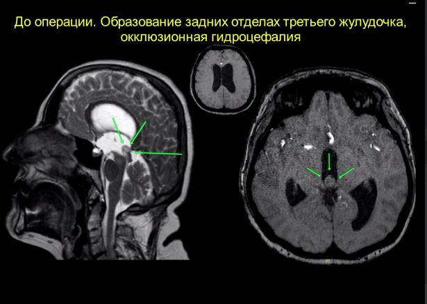
A young patient presented to the department with severe symptoms of intracranial hypertension and impaired motor coordination. A comprehensive examination, including computed tomography (CT), magnetic resonance imaging (MRI), and ophthalmoscopy, revealed a large tumor in the posterior third ventricle with signs of obstruction of the aqueduct of Sylvius and the development of occlusive hydrocephalus. The patient also exhibited signs of intracranial hypertension, including papilledema.
Taking into account the localization of the tumor, rapid progression of clinical symptoms and high risk of decompensation, a decision was made to perform surgical intervention using a complex transcallio-transvenricular-transchoroidal approach to the posterior part of the third ventricle.
During the operation, the necessary instruments were used to control and completely resect the space-occupying lesion.
According to the neurosurgery surgeons, the operation began with a limited craniotomy in the right anterior parasagittal region. Using intraoperative ultrasound, transcallosal access to the corpus callosum was achieved. The trajectory was then directed to the right lateral ventricle, followed by entry into the third ventricle via the sub- and transchoroidal pathways between the choroid plexus and the deep cerebral vein.
In the posterior portion of the third ventricle, near the aqueduct of Sylvius, surgeons discovered a pathological grayish-colored mass obstructing the cerebrospinal fluid (CSF) circulation. The tumor, presumably glial, was completely removed. Endoscopic examination of the original growth zone confirmed the absence of residual tissue.
The operation was performed using a modern surgical microscope, which allows for changing viewing angles, ensuring high precision and controlling parameters during all stages of the intervention.
The patient's condition is currently stable, and all pre-operative complaints have completely resolved. A repeat MRI of the brain with contrast revealed no residual tumor tissue, and the cerebrospinal fluid (CSF) circulation has been restored. A histological examination is currently underway, which will determine further treatment.
The introduction of yet another new neurosurgical technique in the country demonstrates the attention and ongoing support of the country's top leadership for the healthcare system. Equipping the department with modern equipment, including a surgical microscope, allows Russian specialists to continue implementing and performing the latest surgical techniques at a high professional level.